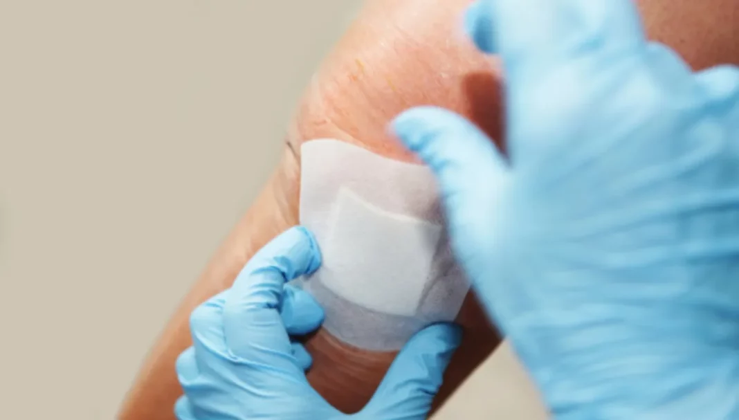
Case Scenario

Mrs Burns, aged 50 years, comes into the pharmacy with a radiation burn that was left open for a week. Hydrocortisone 0.5% with lidocaine 5% ointment and a low-adherent pad was recommended at the time; however, her wound is now quite sore (6/10). Her general practitioner has referred her to see the Wound Care Pharmacist for further advice.

Wound size: 4 x 7 cm

Medical history: Recent radiation therapy for cancer (in remission). No allergies or medicines.

Introduction

Wound infection is a challenging part of wound care management, and systemic antibiotics are commonly prescribed as a treatment of choice for infection.1 However, inappropriate and widespread use of systemic and topical antibiotics are resulting in increased prevalence of methicillin-resistant Staphylococcus aureus (MRSA).1 Chronic wounds affect 2–5% of the population worldwide.2 Accurately identifying the signs and symptoms of wound infection and prompt treatment using evidence-based practice are critical to effective wound infection management,3 and prevention of wound complications and chronic wounds.

Learning objectives

After reading this article, pharmacists should be able to: 

  • Describe the signs and symptoms of a wound infection
  • Discuss strategies to prevent a wound infection
  • Discuss treatment options for a wound infection. 

Competency (2016) standards addressed: 1.1, 1.4, 1.5, 2.2, 3.1, 3.2, 3.5

Accreditation expiry: 30/11/2026

Accreditation number: CAP2312DMLS

Already read the CPD in the journal? Scroll to the bottom to SUBMIT ANSWERS.

Acute and chronic wounds

Wounds that follow wound healing phases within a timely and orderly manner, generally within 2–4 weeks, are described as ‘acute’ (e.g. cuts, grazes, burns, skin tears and postoperative wounds).4

Wounds that do not progress normally through these phases, often due to external and internal factors such as age, comorbidities, medicines, smoking and alcohol, are considered ‘slow to heal’ or ‘chronic’ (e.g. venous leg ulcers, diabetic foot ulcers and malignant wounds).4

Identification of wound infection

All open wounds are contaminated or colonised with bacteria, but the numbers present are usually insufficient to affect healing.5 The transition from a non-infected to an infected wound is a gradual process that occurs when the quantity and virulence of the microorganisms in a wound increases to a level that provokes a host immune response locally, systemically or both, and wound healing is impaired.3,6 The term ‘local wound infection’ describes a phase within the International Wound Infection Institute (IWII) Wound Infection Continuum (WIC) or IWII-WIC.3

The IWII-WIC (see Figure 1) is illustrated as:

Contamination: This is the first phase of WIC, where the wound may acquire microorganisms from normal flora from the periwound skin; however, bacteria levels do not proliferate and do not evoke a significant host reaction or delayed healing.7 Healthy red granulation or pink epithelial may be present.

Colonisation: In this phase, bacteria multiply at a limited level, with no significant host reaction or delayed wound healing observed.3,7 Red granulation may be present with mild inflammation and clear exudate.

Local infection: Colonisation progresses to local infection when bacteria multiply and move deeper into wound tissue, eliciting a host reaction leading to signs of local infection and delayed wound healing.3,7 Signs of a local infection typically include erythema, local warmth, swelling, purulent or yellow discharge, wound breakdown and enlargement, new or increasing pain and increasing malodour.3,6

Spreading infection: This is also known as ‘cellulitis’ and can proliferate beyond the wound border (greater than 2 cm of the wound bed) to deeper tissues, muscles and organs.3 Signs and symptoms may include extending erythema, wound breakdown, induration, malaise/lethargy, loss of appetite, and inflammation or swelling of lymph glands.3,6

Systemic infection: This occurs when the impact of the wound infection proliferates throughout the whole body via the vascular or lymphatic system (systemic).3 Signs include sepsis, septic shock, organ failure and even death.6

When are wound cultures indicated?

Local and spreading infection can be difficult to identify when the signs of infection are not evident. Covert (subtle) and overt (clinical) signs of local wound infection may be masked in people who are immunocompromised, such as in the elderly, people with diabetes, or people who have poor vascular perfusion.3,6 In these people, wound cultures may be warranted when there remains a high clinical suspicion of wound infection, even in the absence of clinical signs and symptoms.3

When is antibiotic use appropriate?

Both topical and systemic antibiotics have the capacity to destroy or inhibit bacterial growth and therefore have a role in managing wound infection. However, it is important that their use be limited to only when necessary, due to increasing concerns regarding microbial resistance.3

In practice, antibiotic administration is often empirical, without the support of microbiological analysis.8 The widespread systemic and topical use of antibiotics fosters the development of resistant bacterial strains or ‘superbugs’ such as MRSA.1,9,10 Antimicrobial resistance (AMR) is a global crisis, and judicious use of antibiotics is critical.6

The use of systemic and topical antibiotics is not appropriate for the routine treatment of colonised or locally infected wounds.1,9 Antibiotics are only appropriate when there are clinical signs of spreading or systemic infection, based on the identified infecting organism.3 Studies suggest that there is excessive use of antibiotics in individuals with non-healing wounds.10 There is also evidence that the routine use of antibiotics in clinically infected leg ulcers has no benefit and can be harmful due to the colonisation of resistant organisms.1

Topical antibiotics do not penetrate wound biofilms and deep skin infections and are not appropriate for the management of local wound bacterial burden.1,11 Risks with use include evoking hypersensitivity reactions, contributing to antibiotic resistance, and can potentially cause systemic absorption when applied to large wounds.11,12

What is a wound biofilm?

A biofilm is a cluster or community of bacterial cells embedded in an extracellular polysaccharide (EPS) matrix that acts as a barrier, rendering it resistant to the immune system, as well as many antiseptics and antibiotics.7,13 Biofilms are not visible to the naked eye and cannot be detected by routine swabbing.13 Biofilms have been found to be present in over 90% of chronic wounds.14 Biofilms are suspected if a wound fails to respond to appropriate systemic antibiotics or antiseptics or fails to heal at the expected rate with optimal care.6,13

The concept of wound biofilm management is a method to manage infection. This requires good wound bed preparation through regular debridement, combined with the use of topical antimicrobials that can penetrate the EPS and attack bacteria within the matrix, aiding in the removal and suppression of biofilms.6,14

Prevention of infection

Wound hygiene

Having a competent immune system combined with good wound care practice can reduce the risk of a wound becoming infected. Wound hygiene using wound cleansing and debridement techniques can reduce bacterial load.7,10 For most acute wounds, cleaning away wound debris, excess exudate, loose slough, and dressing residue using saline is recommended to reduce bacterial load to an acceptable level.9,12

Debriding a wound is the removal of non-viable tissue, foreign material, senescent cells and bacteria, which can aid in the suppression and removal of biofilms.14 For infected wounds with higher bacterial load, such as purulent or ‘sloughy’ wounds, and wounds with suspected biofilms, a range of non-toxic antiseptics is usually sufficient to manage local infection without the need for antibiotics.7

The IWII Wound Infection Continuum and Management Guide illustrates a holistic approach to assessment and management of the individual and their wound. This includes guidance on identification of wound infection and potential biofilm, selection of cleaning solution, debridement of the wound, and choice of wound dressing.10 A copy of this guide is available on the IWII website: https://woundinfection-institute.com/wp-content/uploads/IWII-WIC-A3-1.pdf

The role of dressings

Keeping wounds covered in a moist healing environment plays a key role in preventing wound complications such as infections. Occlusive dressings have been shown to provide a moist environment that assists with the reepithelialisation of cells15,16 and allows epithelial cells and fibroblasts to migrate more easily through the fluid to achieve optimum healing.16 In contrast, when a wound is left open to dry out, cells dehydrate which causes a scab or crust to form over the wound.17 Essentially the scab acts as a physical barrier to the movement of cells across the wound bed, and reepithelialisation and wound healing become inhibited.16,17

Management of wound infections

Antiseptic treatment options

An antiseptic is a type of ‘topical antimicrobial’ that inhibits and kills microorganisms present within a wound or on intact skin.18 Antiseptics are generally non-selective and have a broad spectrum of antibacterial activity,6 and the risk of bacterial resistance is often low due to their action at multiple sites within microbial cells.7,18 Topical antimicrobials play a role in preventing and managing wound infection, and are used in the treatment of clinically infected open acute and chronic wounds.3,7 Antiseptics should also be used in conjunction with systemic antibiotics in the presence of spreading or systemic infection.7

Judicious use of antiseptics is essential, as some antiseptics are considered ‘cytotoxic’ and can kill healthy wound tissue.3,6 Cytotoxicity of antiseptics is thought to be concentration dependent, and antiseptic use is recommended at the lowest concentration to reduce tissue damage.6

There are several topical antiseptics appropriate and safe for use in local wound infections, including medical-grade honey, silver dressings (in ionic and nanocrystalline forms), and cadexomer-iodine formulations.3,11,18,19 Evidence has shown that in superficial burns, manuka honey had higher complete healing rates than silver sulfadiazine and topical antibiotics.3 

Antiseptics commonly used in wound infection management

  • Manuka honey (medical grade)
    • Example: Medihoney wound gel, Manukalife WoundGel 800+, Dermagen Manuka Oil Balm
    • Available as amorphous gel or impregnated dressings18
    • Medical-grade honey derived from the Leptospermum species
    • Effective against Gram-positive and Gram-negative bacteria, including Escherichia coli, Pseudomonas aeruginosa, Staphylococcus aureus, Acinetobacter, and MRSA3,19
    • Antibacterial, anti-inflammatory, antifungal
    • Provides autolytic debridement and inhibits biofilm formation.19
  • Silver (elemental, organic)
    • Examples: Atrauman Ag, Acticoat Ag, Aquacel Ag
    • Available as impregnated dressings3,18 with sustained release of silver
    • Silver sulfadiazine (SSD) in cream form is short-acting, requires reapplication at least daily, and is no longer recommended20
    • Broad-spectrum activity against Gram-negative and Gram-positive bacteria, including P. aeruginosa, E. coli, and S. aureus3
    • Inhibits biofilm formation18
    • No to mild concentration-dependent cytotoxic effect on fibroblasts3
    • Limit to 2 weeks of use and up to 4 weeks if the wound is showing improvement.
  • Cadexomer iodine 0.9%
    • Examples: Iodosorb ointment, powder, tulle
    • Available as an ointment, powder, paste, and impregnated dressings18
    • Broad-spectrum activity against Gram-negative and Gram-positive bacteria, fungi, spores, protozoa, and viruses3
    • Provides autolytic debridement and desloughing action13
    • Reduces microbial burden complicated by biofilm at 0.9% concentration3,13
    • Improved wound healing when compared with other dressings such as hydrocolloid, paraffin gauze, non-adhesive dressing, and saline wet-to-dry dressings21
    • Contraindicated in children younger than 12 years, iodine sensitivity, thyroid or renal disorders, and large burns.3
  • Povidone-iodine 10%
    • Examples: Betadine liquid, ointment, cream, Inadine tulle
    • Available as an ointment, liquid gel, and impregnated dressing18
    • Dose-dependent cytotoxic effect on fibroblasts3,13
    • A study showed that human fibroblast growth was completely inhibited even at lower concentrations of 0.1% and 1% povidone-iodine solutions22
    • Contraindicated in neonates, iodine sensitivity, thyroid or renal disorders, and large burns.3

The pharmacist’s role in wound care

Pharmacists play an important role in the identification, prevention and management of wound care, contributing significantly to patient wellbeing. This can be done by educating patients on how to identify signs and symptoms of wound infection.

Pharmacists are also in a position to assess the severity of a patient’s wound, their infection risk and overall patient health, which can aid in early detection and appropriate referral as needed.

When advising on wound care, pharmacists can offer valuable guidance on preventive measures to reduce the risk of wound infection by educating patients on proper wound care techniques, hygiene practices, and the role of different wound dressings and the moist wound healing concept.

A study in Australia showed that patients were satisfied with their experience with acute wound care at pharmacy-based wound clinics, and felt that receiving information on their progress of wound healing and how to self-manage wounds was beneficial.23 Pharmacists can contribute to antimicrobial stewardship (AMS) by recommending antimicrobials only in wounds with clinical signs and symptoms of infection, and referring patients with suspected wound infection due to a compromised immune system.6

Conclusion

Healthcare professionals need to ensure that their practice in the identification, prevention and management of wound infection is consistent with evidence-based guidelines and AMS.

It is essential to provide a holistic assessment of patients and their wounds, involve patients in their own care, and ensure that they are at the centre of the multidisciplinary team. Pharmacists can empower patients using clear communication and providing education to patients, to optimise the management of wound infection, enhance self-care skills and improve clinical outcomes.

Case scenario continued

Day 1:

On assessment, the wound appears locally infected with signs of pain, redness, and yellow pustules (see Day 1); however, there are no signs of spreading or systemic infection. As per IWII-IWC, local infection requires a topical antimicrobial to control the wound burden, therefore an alternative treatment plan is provided. 

The wound is cleansed with saline and gauze. A silver-impregnated tulle dressing is applied as a primary dressing, and a silicone foam border dressing is applied as a water-resistant secondary dressing. Mrs. Burns is advised to keep the dressing on for 3 days, to report any signs of increasing pain or discharge, and to come back for review.

wound infectionDay 3:

Mrs. Burns reports that the wound is no longer sore, and that it felt comfortable soon after having it dressed and covered. 

The dressing is removed, and the wound bed is now pinkish-red with new epithelial tissue, and yellow pustules are no longer visible (see Day 3). The wound is cleansed, and a silicone foam border dressing is used to protect it for another 4–5 days until the wound has fully closed.

wound infectionDay 7:

Mrs. Burns presents with a wound that has closed with healthy new epithelial tissue present (see Day 7). She is advised to keep it hydrated by moisturising twice daily with an emollient to strengthen new tissue.

wound infection

Reproduced with permission from L Sheehan. Images provided with consent from patient for educational purposes.


Key points

  • Keeping wounds clean and covered in a moist healing environment using appropriate dressings is key to preventing wound complications such as infections.
  • Various topical antiseptics are appropriate and safe for use in local wound infections, including medical-grade honey, silver dressings, and slow-release iodine formulations.
  • Use of systemic and topical antibiotics is not appropriate for the routine treatment of colonized or infected wounds.1,9
  • Antibiotics are only appropriate when there are clinical signs of a spreading or systemic infection,3 and may be required in patients who are immunocompromised, based on the identified infecting organism.

This article is accredited for group 2 CPD credits. Click submit answers to complete the quiz and automatically record CPD against your record.

SUBMIT ANSWERS

If you do get an enrolment error, please click here

References

  1. White RJ, Cooper R, Kingsley A. Wound colonization and infection: the role of topical antimicrobials. Br J Nurs 2001;10(9):563-78.
  2. Jarbrink K, Ni G, Sonnergren H, et al. The humanistic and economic burden of chronic wounds: a protocol for a systematic review. Syst Rev 2017;6(1):15.
  3. Swanson T, Ousey K, Haesler E, et al. IWII wound infection in clinical practice consensus document: 2022 update. J Wound Care 2022;31(Sup12):S10-S21.
  4. Stacey M. Why don’t wounds heal? Wounds International 2016;7:16-21.
  5. Hayward P, Morrison W. Current concepts in wound dressings. Aust Prescr 1996;19:3-11.
  6. Haesler E, Ousey K. Evolution of the wound infection continuum. Wounds International 2018;9:6-10.
  7. Wound infection in clinical practice. An international consensus. Int Wound J 2008;5 Suppl 3(Suppl 3):iii-11.
  8. Puca V, Marulli RZ, Grande R, et al. Microbial species isolated from infected wounds and antimicrobial resistance analysis: data emerging from a three-years retrospective study. Antibiotics (Basel) 2021;10(10).
  9. White RJ, Cutting K, Kingsley A. Topical antimicrobials in the control of wound bioburden. Ostomy Wound Manage 2006;52(8):26-58.
  10. International wound infection institute. IWII wound infection continuum and management guide. 2022 At: https://woundinfection-institute.com/wp-content/uploads/IWII-WIC-A3-1.pdf
  11. Ousey K, Swanson T, Sussman G. Wound infection in clinical practice – made easy. Wounds International. 2022.
  12. Healy B, Freedman A. ABC of wound healing: infections. BMJ 2006;332:838-41.
  13. Wounds UK. Best Practice Statement. The use of tropical antimicrobial agents in wound management. 3rd edn. London: Wounds UK, 2013.
  14. Sandoz H, Swanson T, Weir D, et al. Biofilm-based wound care with cadexomer iodine – made easy. Wounds International. 2017.
  15. Attinger C, Wolcott R. Clinically addressing biofilm in chronic wounds. Adv Wound Care (New Rochelle) 2012;1(3):127-32.
  16. Brennan SS, Foster ME, Leaper DJ. Antiseptic toxicity in wounds healing by secondary intention. Journal of Hospital Infection 1986;8(3):263-7.
  17. Rheinecker SB. Wound management: the occlusive dressing. J Athl Train 1995;30(2):143-6.
  18. Tan ST, Dosan R. Lessons from epithelialization: the reason behind moist wound environment. The Open Dermatology Journal 2019;13:34-40.
  19. Vowden P, Vowden K, Carville K. Antimicrobial dressings – made easy. Wounds International 2011;2(1):1-6.
  20. Maillard JY, Kampf G, Cooper R. Antimicrobial stewardship of antiseptics that are pertinent to wounds: the need for a united approach. JAC Antimicrob Resist 2021;3(1):dlab027.
  21. Leaper D. Appropriate use of silver dressings in wounds: international consensus document. Int Wound J 2012;9(5):461-464.
  22. Miller CN, Newall N, Kapp SE, et al. A randomized-controlled trial comparing cadexomer iodine and nanocrystalline silver on the healing of leg ulcers. Wound Repair Regen 2010;18(4):359-67.
  23. Balin AK, Pratt L. Dilute povidone-iodine solutions inhibit human skin fibroblast growth. Dermatol Surg 2002;28(3):210-4.
  24. Sheehan L, Dias S, Joseph M, et al. Primary care wound clinics: a qualitative descriptive study of patient experiences in community pharmacies. Pharmacy (Basel) 2022;10(4).

Our author

Lusi Sheehan (she/her) BPharm, GradCertWoundCare, AACPA, DipMgmt, ADvPP(II), MPS is a credentialed Wound Care Clinician, an Accredited HMR Pharmacist and a Professional Services Pharmacist. Known as ‘The Wound Educator’, she has a postgraduate degree in Wound Care from Monash University and provides wound care education to pharmacists and allied health professionals Australia-wide.

Our reviewer

Daisy Cheung (she/her) BPharm, MPS

Conflict of interest declaration

Lusi runs her own wound care education course for pharmacists Australia-wide.